Gallery
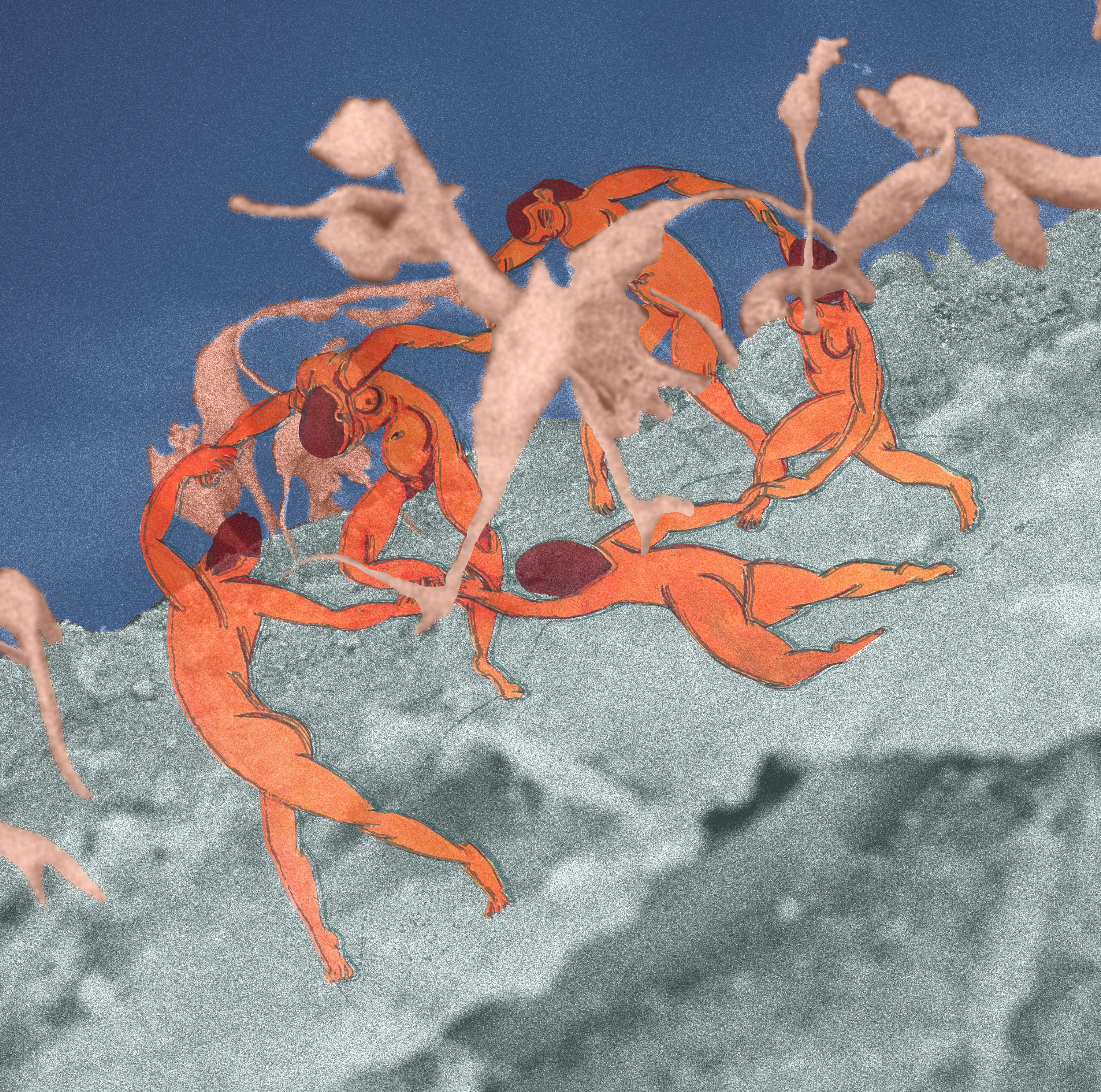
Dr. Vinnacombe-Willson has interests in scientific artwork. She worked as an illustrator for the book “Playing with Math: Stories from Math Circles, Homeschoolers, and Passionate Teachers” in 2012. Now she primarily works using a combination of digitally hand-drawn images, Inkscape, and Blender. For the latter, she completed two courses in 2023 and 2024. She has also participated in various scientific image contests and enjoys sample preparation and characterization of all kinds of materials using electron microscopy.
Covers

Image Contest Entries


Electron Microscopy Image Gallery
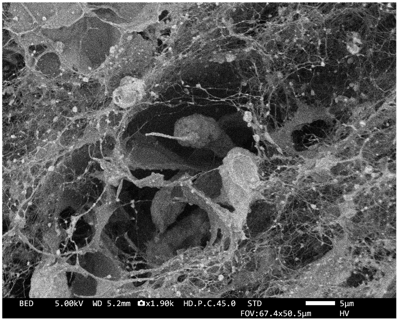
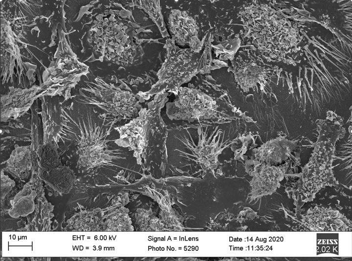
 Cancer cells in dECM
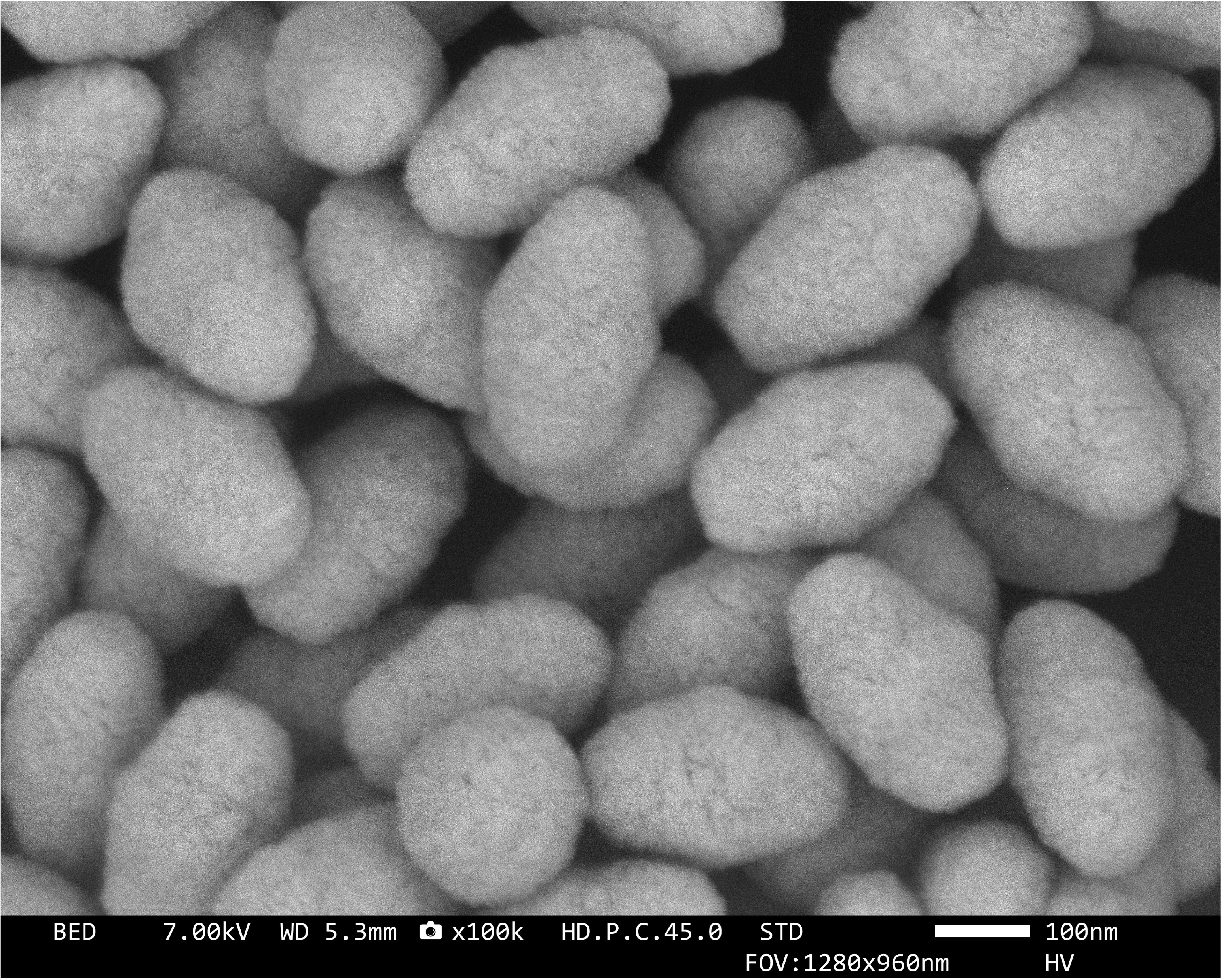
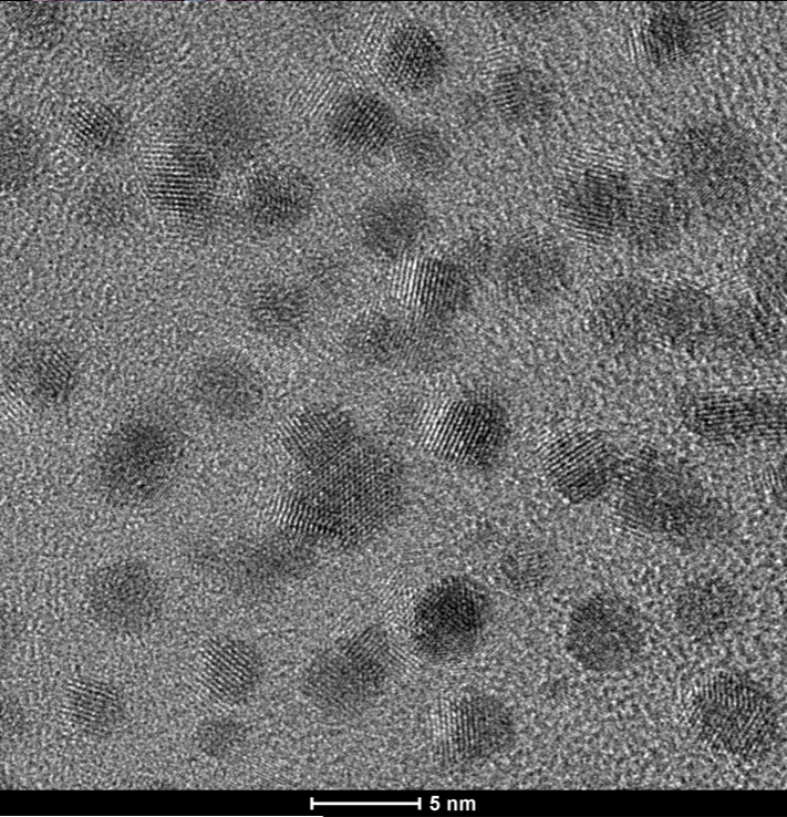
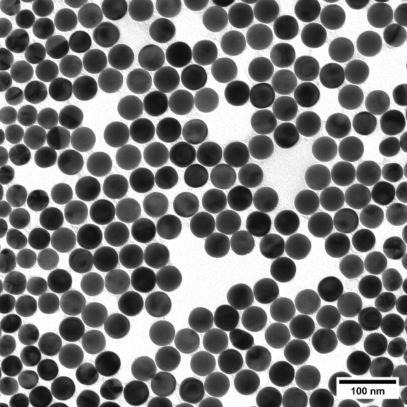 Gold nanospheres
 Salt crystals on gold nanostars
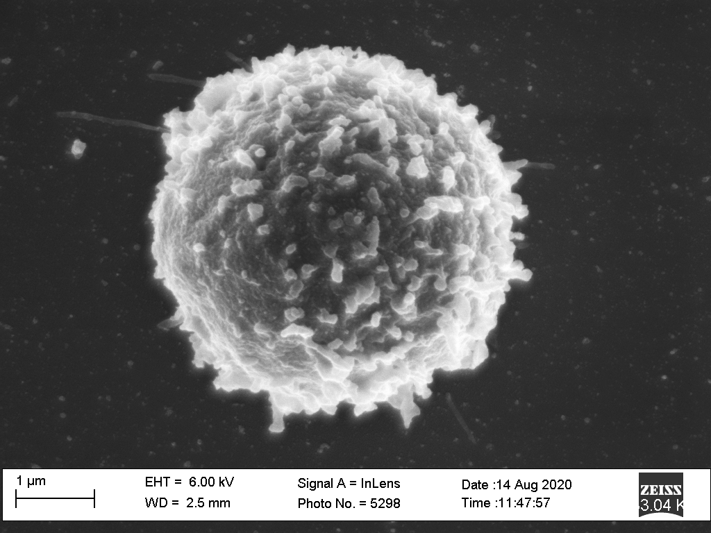 T-cell
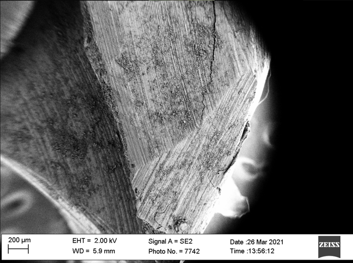 Human Tooth
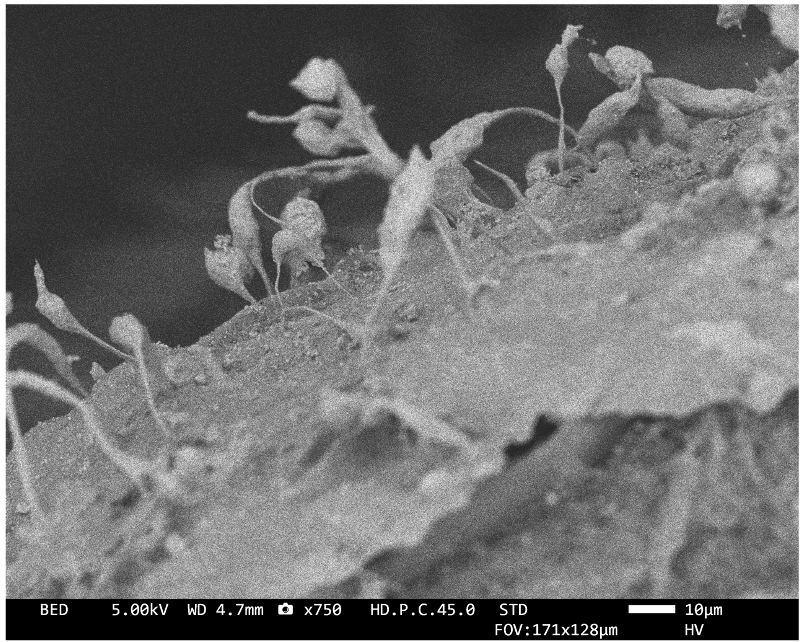
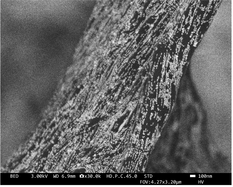
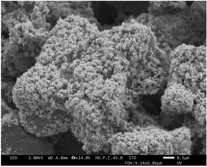
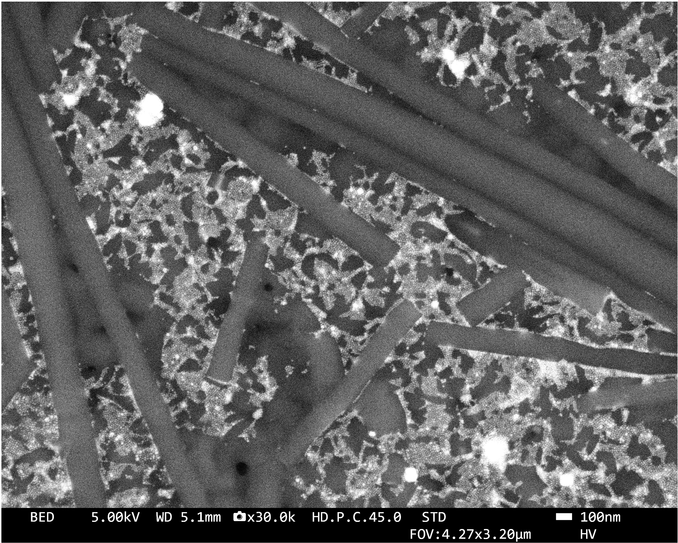 Perovskite nanocrystals on organic nanotubes
 SH-SY5Y neuroblastoma cells on gold nanostars
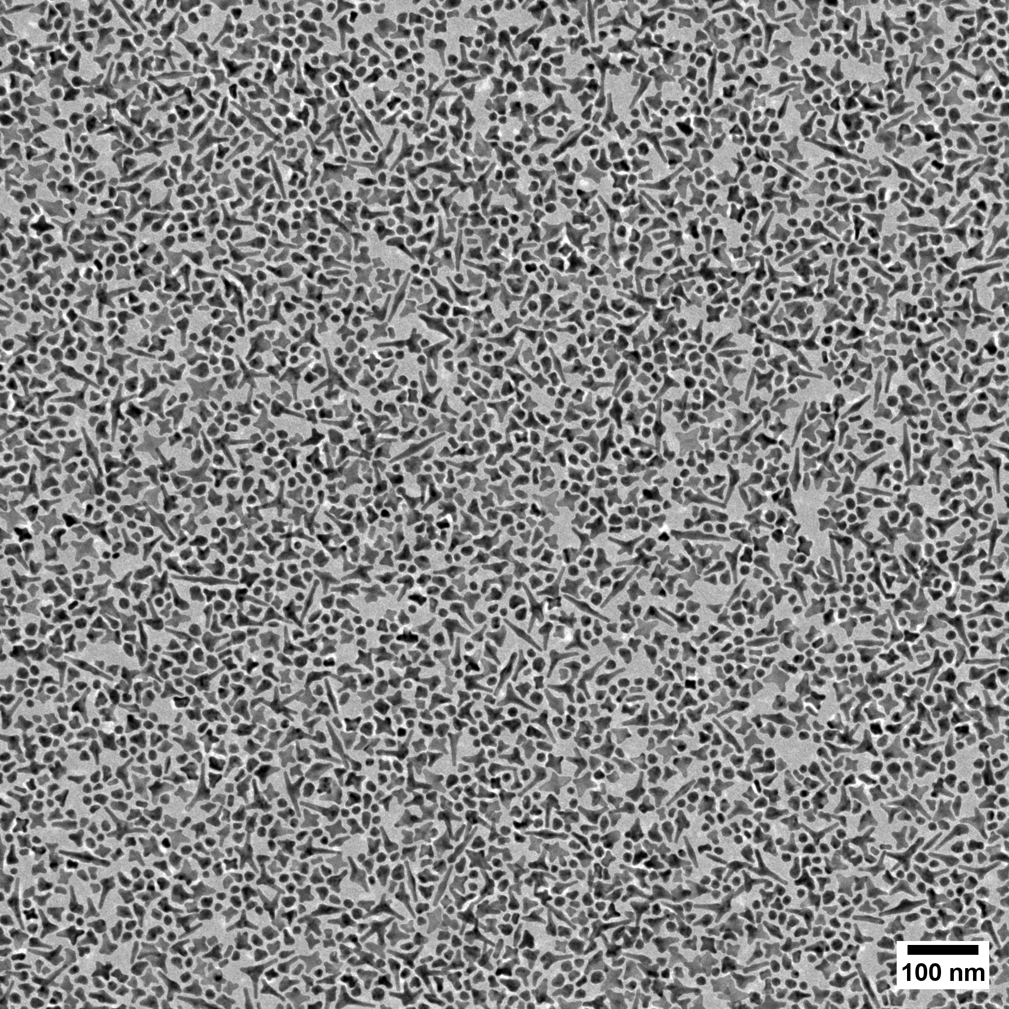
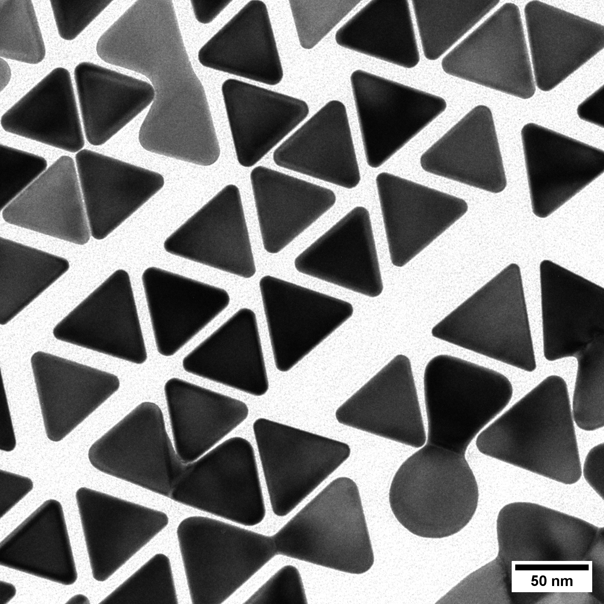 Gold nanotriangles
 MDA-MB-231 breast cancer cells in dECM
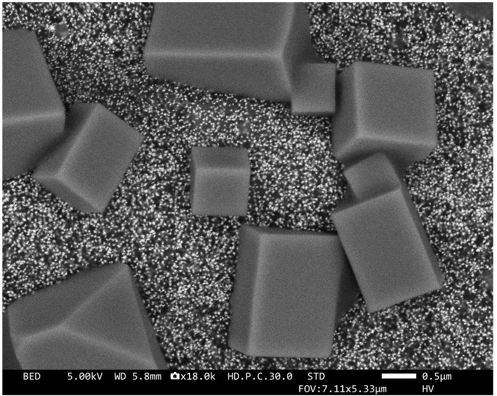
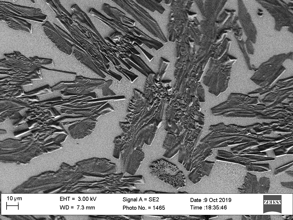 KCN crystals on a gold substrate
 Gold nanorattles
 Chiral gold bipyramids
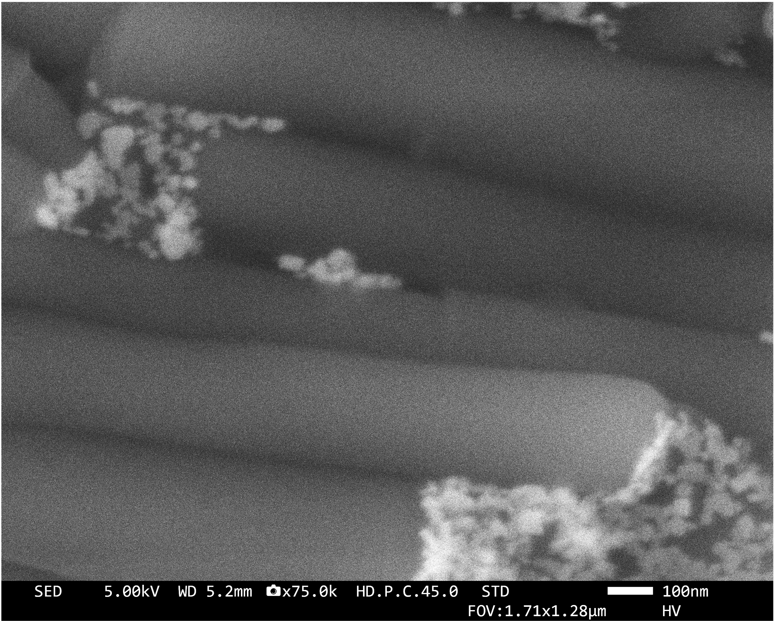 Perovskite nanocrystals on organic nanotubes close up
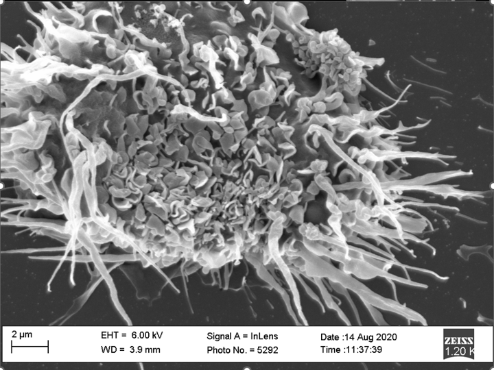 Dendritic cell
 Gold nanorods on standing organic wires
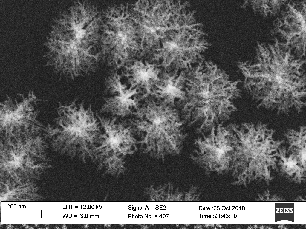
 Gold nanostars
 Dendritic cells
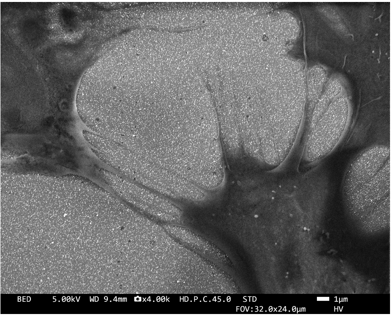
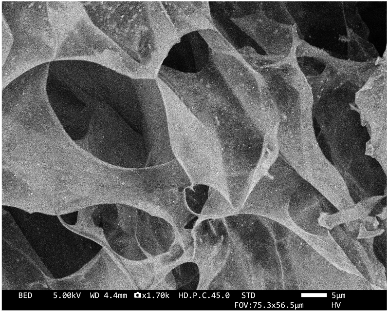
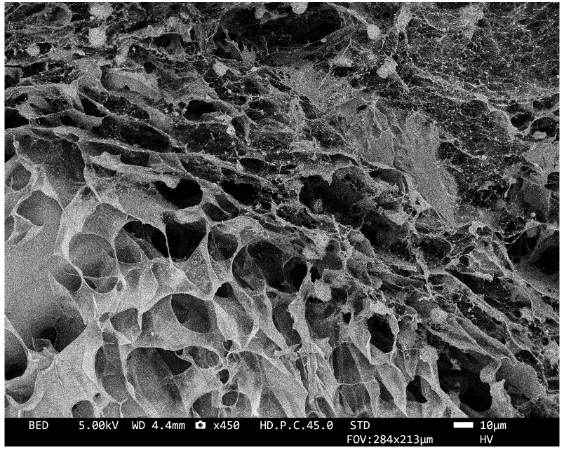 Decellularized extracellular matrix
 Small gold nanospheres
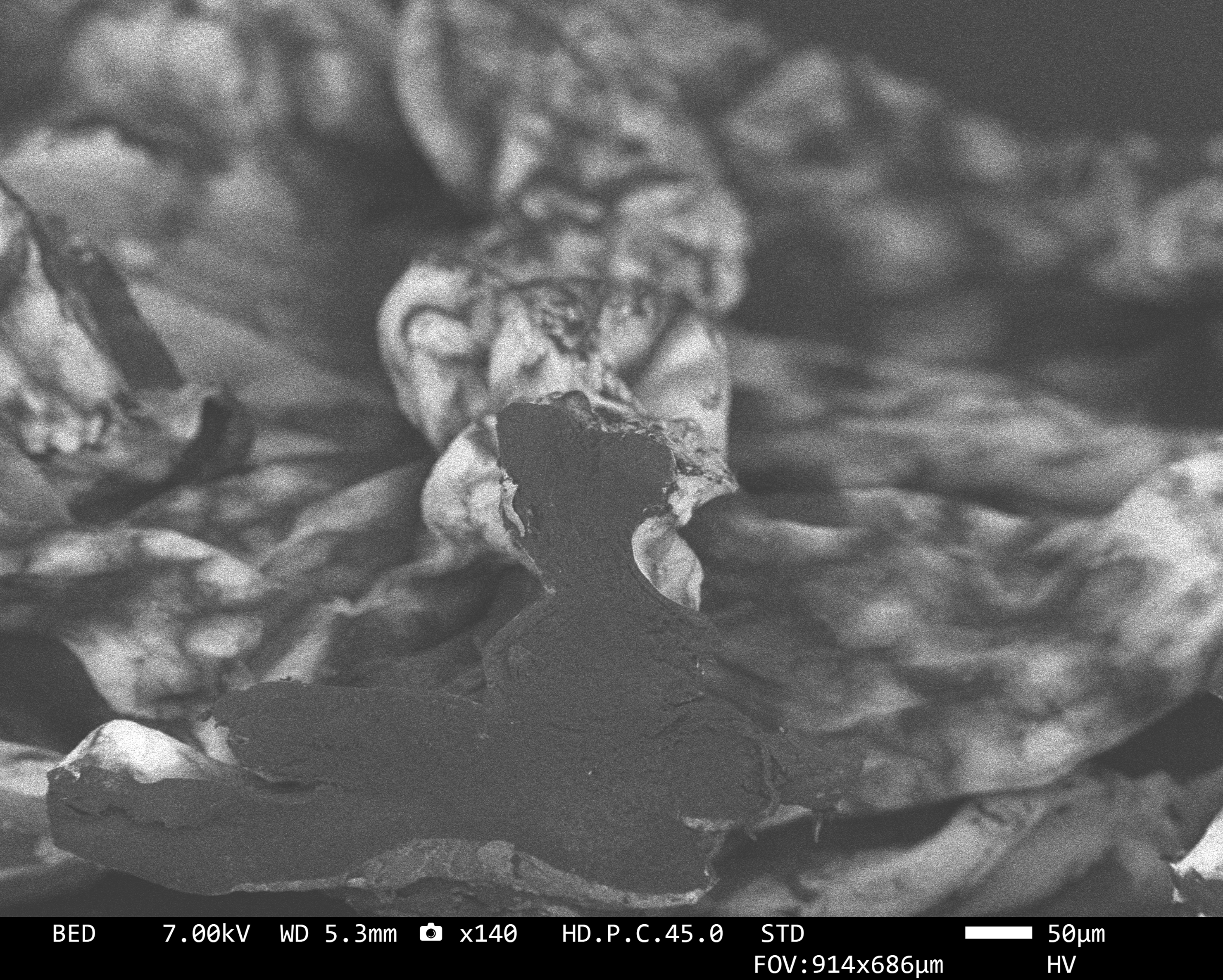
 BiVO4 microparticle with gold nanostars
 Hydrogel containing gold nanorods
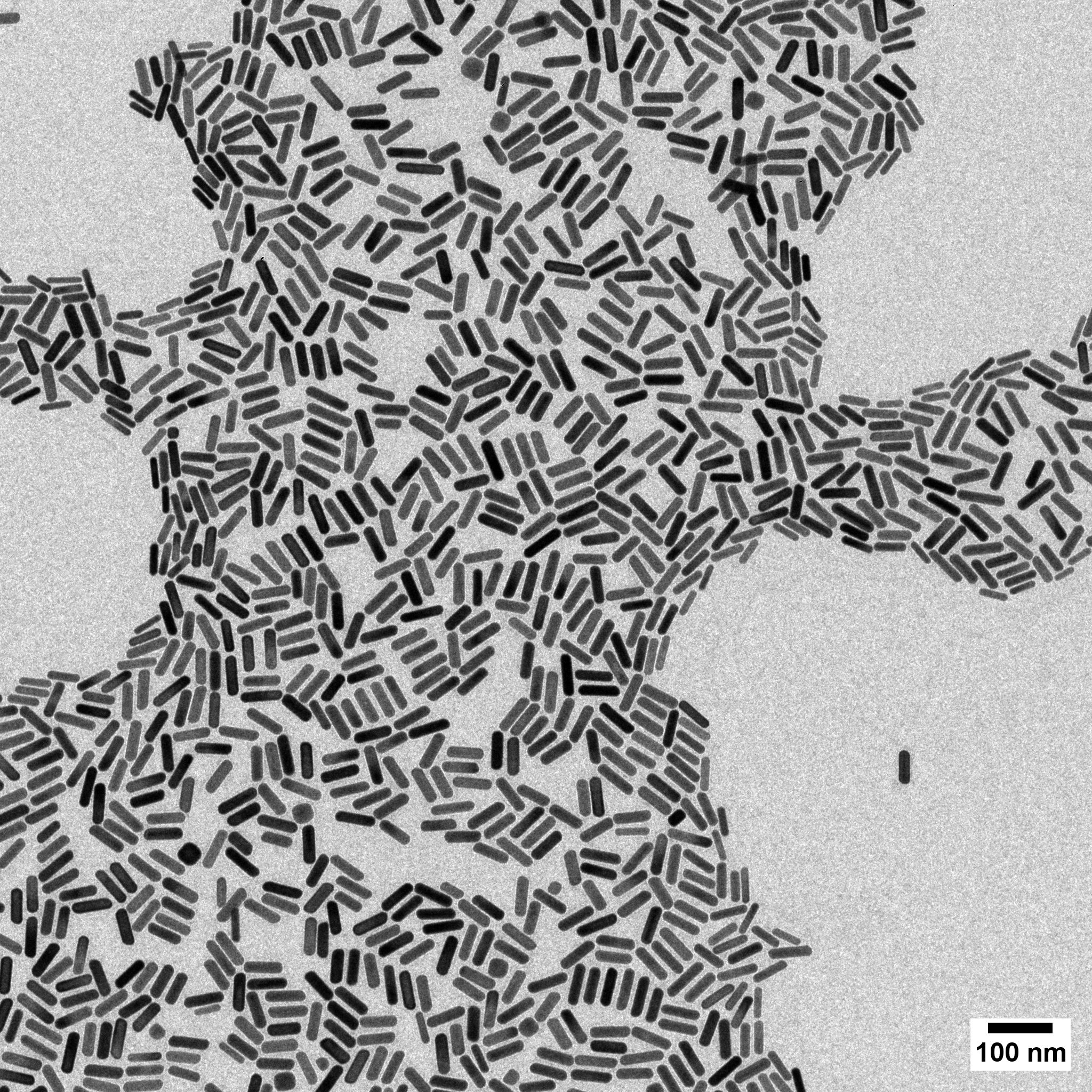 Gold nanorods
 Dendritic gold nanoparticles grown on glass
 Hydrogel cell culture scaffold with a gold nanostar coating
×
❮
![Expanded Image]() ❯
❯